






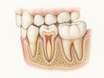




If you feel a hard, sharp bump in your gum after a dental procedure, it is usually a bone spicule—a small piece of bone working its way through the tissue as the area heals. Most spicules are not a sign that your dental work failed, and many either come out on their own or can be removed quickly by a dentist. Keep the area clean, stick with soft foods, and do not pick at the spot. Call your dentist if you have worsening pain, swelling, pus, fever, trouble swallowing, or if it is still quite sore after 1–2 weeks.
Do not try to dig out a bone spur with tweezers, a needle, or your fingernail. That can turn a small healing bump into a real infection.
Why It Happens After Dental Work
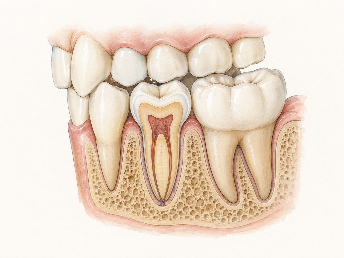
A bone spicule is a tiny piece of bone that can move upward through the gum as your mouth heals. It can happen after a tooth extraction, dental implant placement, a deep cleaning, or a mouth injury. During these procedures, bone and gum tissue can be disturbed, and small fragments may be left behind or loosen near the surface.
This usually is not a sign that the dentist made a mistake. In a study of bone spicules around dental implants, tiny bone fragments showed up in soft tissue and did not appear to cause major inflammation. The report described them as a common, often unrecognized finding after implant site preparation. PMC study on bony spicules around implants
Sometimes the bump is not new bone but a retained root fragment—a leftover piece of tooth root. Other times it comes from natural bone reshaping, called bone remodeling. A review of mouth ulcers with exposed bone found that events like tooth removal, deep scaling, and even long-term pressure from chewing or brushing could be involved. Narrative systematic review on oral ulceration with bone sequestration
Bone Spur vs. Other Gum Bumps
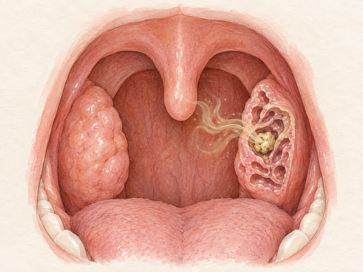
A typical bone spur feels hard and sharp. You may see a small white, yellow, or gray speck where the tip is close to the surface. It can be tender when food or your tongue presses against it.
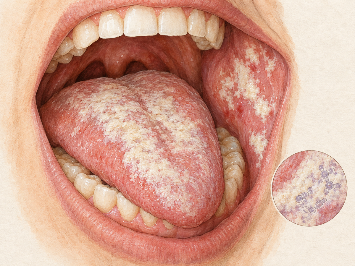
Other gum bumps can look similar but feel and behave differently:
- A canker sore is a shallow mouth ulcer. It is soft, painful, and usually white or yellow with a red edge.
- A dental abscess is a pus-filled infection. The bump may feel squishy, warm, or tender, and you may have a bad taste or fever.
- An oral cyst is a fluid-filled sac. It can be firm or soft, sometimes with a bluish color.
- Periodontal disease, or gum disease, can cause redness, swelling, bleeding, and tenderness, but not one hard, sharp splinter.
Many gum lumps overlap in appearance, so a dental exam is the safest way to know what you are dealing with. Lumps and bumps of the gingiva Do not poke or press the spot with dirty tools or fingers.
Safe Home Care for Relief
Home care is meant to calm the area while the spicule works out or until a dentist removes it. It will not force the bone out, but it can make the wait more comfortable.
- Rinse with warm salt water 2–3 times a day. Use about half a teaspoon of salt in a cup of warm water and swish gently. Salt-water rinsing after tooth removal has been linked to fewer post-operative complications in a clinical trial. Salt water mouthwash after extraction A review also found warm saline mouth baths may help reduce complications, though the authors called for more study. Warm saline mouth bath review
- Brush gently around the area. Keep the rest of your mouth clean, but avoid scrubbing the bump itself.
- Eat a soft food diet until the area settles. Yogurt, soup, mashed potatoes, eggs, and smoothies are easier on the gum.
- Use a cold compress on the outside of the cheek if there is swelling. A cloth-covered ice pack for 10–15 minutes at a time can help.
- Ask a dentist or pharmacist whether over-the-counter pain relief such as acetaminophen or ibuprofen is safe for you.
For similar mouth sores with exposed bone, conservative care with cleaning and pain relief may be used while the body separates the loose fragment or until removal is possible. Review on oral ulceration with bone sequestration
When to Call Your Dentist
Call your dentist promptly if you notice any of these:
- Pain that keeps getting worse
- Swelling that is spreading
- Pus, a bad taste, or a bad smell from the area
- Fever or feeling generally unwell
- Bleeding that does not stop
- Trouble swallowing, opening your mouth, or breathing
- A spur that is still very tender or has not improved after 1–2 weeks
These can be signs of infection or a retained piece that needs professional care. In a study of delayed infections after wisdom-tooth removal, swelling and pus were the main signs of infection that showed up weeks later. Delayed-onset infections after extractions Dental infections can become serious, so difficulty swallowing or breathing should be treated as an emergency. Guidance on dental infection management
If you are anxious, have a medical condition that slows healing, or take medicines that affect your bones or immune system, call sooner rather than later.
In-Office Removal and Treatment
When you visit, the dentist will usually:
- Examine the bump and ask how it has felt.
- Take a dental X-ray if needed to check for a root fragment or sharp bone edge.
- Numb the area with local anesthesia if removal is needed.
- Remove the spicule with sterile instruments through a tiny opening or after gently loosening the gum.
- Smooth any sharp bone edges so the gum can close and heal.
- Prescribe antibiotics if there is an infection.
- Monitor the spot if the spicule is small, loose, and likely to come out on its own.
A review of oral ulcers with exposed bone found that removing the loose, non-living bone fragment was a common and effective treatment, with most cases healing within a few weeks. Review on oral ulceration with bone sequestration The procedure is usually quick, and many people feel relief soon after the sharp edge is gone.
Healing Timeline and Prevention
Most small bone spicules improve within days to a few weeks. Bigger or deeper fragments can take longer, especially if food or dentures keep pressing on the spot. In a review of similar mouth sores with exposed bone, many lesions had been present for two weeks or less, and the majority healed within 4 weeks after the loose bone was removed. Review on oral ulceration with bone sequestration
To support healing:
- Follow the aftercare directions you received after any dental surgery.
- Keep your mouth clean, but be gentle near the bump.
- Avoid hard, crunchy, or sharp foods that can push the spur further into the gum.
- Go to any follow-up appointments so the dentist can check the area.
Not every spicule can be prevented. Bone remodeling after an extraction or implant means the body is reshaping the bone, and small fragments can surface even with careful treatment. That is part of how the body sometimes heals, not a personal failure. Bony spicules around implants If the bump is painful, persistent, or worrying you, a quick dental visit is the simplest next step.
This article has been reviewed by an oral health professional for accuracy. It is intended to provide general educational information and should not be used as a substitute for personalized medical advice, diagnosis, or treatment. If you have questions or concerns about your oral health, please consult a dentist, physician, or other qualified healthcare provider.